In vivo animal models are an important tool for the understanding of human development and disease. Studies using the frog Xenopus have made remarkable contributions to our understanding of fundamental processes such as cell cycle regulation, transcription, translation and many other topics. Xenopus is remarkable for studying development and disease, including birth defects, cancer, and stem cell biology. Because Xenopus are easy to raise, producing many thousands of eggs per day, these frogs have emerged as a premiere model for understanding of human biology from the fundamental building blocks to the whole organism.
The recent development of CRISPR/Cas9 technology has made it easy to target genes of interest using Xenopus.This course has been designed with that in mind. Our goal is for each student to design a set of experiments focusing on their gene or biological interest. Prior to starting the course, students will be expected to choose gene(s) of interest, and the instructors will generate sgRNAs targeting these genes. These can be the students’ own genes, or chosen from a bank provided by the instructors.
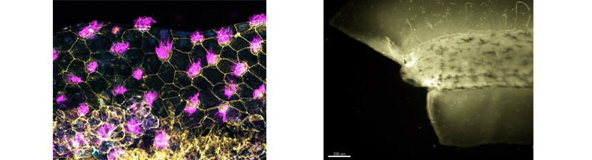
During the course, the students will analyze any phenotypes generated from CRISPR/Cas9based gene depletion while learning the diverse array of techniques available in Xenopus. In previous courses, we have guided students in the ablation of a wide variety of genes and helped them design suitable assays for their biological interests. Most recently, students have targeted autism genes, thyroid genes and immune modulators, several of which have already led to publications. Approaches covered will include microinjection and molecular manipulations such as CRISPR/Cas9 knockouts, antisense morpholino-based depletions, transgenics, and mRNA overexpression. In addition, students can combine these techniques with explant and transplant methods to simplify or test tissue level interactions. Additional methods include mRNA in situ hybridization and protein immunohistochemistry as well as basic bioinformatic techniques for gene comparison and functional analysis. Biochemical approaches such as proteomics and mass spectrometry and biomechanical concepts will also be discussed. Finally, to visualize subcellular and intercellular activities, we will introduce a variety of sample preparation and imaging methods including time-lapse, fluorescent imaging, optical coherence tomography and confocal microscopy. These are facilitated by state-of-the-art equipment from Nikon, Leica, Thorlabs, and Bruker.
Due to the tailored nature of this course, it is suitable for those new to the Xenopus field, as well as for more advanced students who are interested in emerging technologies. Please feel free to contact the instructors for informal guidance.
2019 Lecturers:
Sang-Wook Cha, University of Central Missouri
Ken Cho, University of California Irvine
Frank Conlon, University of North Carolina
Rebecca Heald, University of California, Berkeley
Raymond Keller, University of Virginia
Darcey Kelley, Columbia University
Roberto Mayor, University College London, UK
Nanette Nascone-Yoder, North Carolina State University
Andrea Wills, University of Washington
Sarah Woolner, University of Manchester, UK
10fps of Xenopus GRP cilia
10 frames per second imaging of Xenopus GRP cilia. GFP labeled cilia, RFP membrane marker. Courtesy of Melanie Tingler, Shiaulou Yuan, and Mustafa Khokha. Cold Spring Harbor Xenopus Course, Yale University. Movie generated on Bruker Opterra II confocal microscope.
125 fps blood flow
125 frames per second imaging of Xenopus red blood cells in gills. Playback at 5x slow motion. Video courtesy of Vaughn Colleluori and Mustafa Khokha, Cold Spring Harbor Xenopus Course,Yale University. Movie generated on Bruker Opterra II confocal microscope.
CLAMP GFP and RFP
CLAMP GFP (green) marks tips of cilia and membrane RFP (red) marks cilia axoneme. Two color images collected at 20 fps. CSHL 2015 Xenopus course. Movie generated on Bruker Opterra II confocal microscope.
Support & Stipends
Stipends are available to offset tuition costs as follows:
Please indicate your eligibility for funding in your stipend request submitted when you apply to the course. Stipend requests do not affect selection decisions made by the instructors.


We would like to acknowledge the following companies that provided invaluable support:
Microscopes: Bruker, Morrell Instruments, Nikon Instruments, ThermoFisher Scientific, Thorlabs
Lab Equipment and Software: Bitplane, Electron Microscopy Sciences, Harvard Apparatus, Narishige International USA, Sutter Instrument Company
Discounted Products: Xenopus1
Cost (including board and lodging): $3,960
No fees are due until you have completed the full application process and are accepted into the course. Students accepted into the course should plan to arrive by early evening on April 2 and plan to depart at any time on April 16.
Before applying, ensure you have:
- Personal statement/essay;
- Letter(s) of recommendation;
- Curriculum vitae/resume (optional);
- Financial aid request (optional).
More details.
If you are not ready to fully apply but wish to express interest in applying, receive a reminder two weeks prior to the deadline, and tell us about your financial aid requirements, click below: